
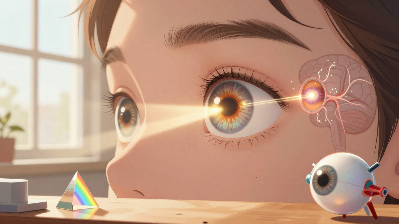
The eye operates as a sophisticated optical system, with every tissue playing an essential part in forming a view of the surrounding world. Through the interaction of curved surfaces and specialised membranes, incoming light is converted each day into signals the brain can understand.
How do the cornea and iris regulate incoming light?
The cornea forms the outermost layer of the eyeball. This transparent, curved structure acts as the first protective lens, bending the initial light rays towards the internal regions and providing the sharpness required to create the correct image.
Immediately behind this first lens sits the iris, the coloured area that dynamically controls the central opening known as the pupil. In bright places, this opening narrows to protect the back of the eye; in dark settings, it widens to collect as much brightness as possible.
Why do coloured lights appear when you press on closed eyes?
When pressure is applied to the eyelids, the mechanical stimulus directly activates the retina's photoreceptors, causing the nervous system to interpret it… Read more
What role does the lens play in focusing images?
Located just behind the pupil, the lens functions as a highly transparent, flexible lens within the eye. Its main task is to continually adjust its curvature, accurately focusing objects at varying distances within the visual field.
When looking at objects very close by, this gelatinous structure becomes thicker in order to converge light rays precisely. If the lens loses its natural transparency over time, a cataract develops, seriously impairing the passage of light to the back of the eye.
Below is a video from the Manual do Mundo YouTube channel that explores the points discussed in this topic in greater depth:
How does the retina convert light into visual signals?
At the back of the eyeball, the retina works much like a photographic sensor filled with sensitive cells. Its reddish, richly supplied surface receives the light projection focused by the lenses, beginning the decoding of patterns in the external environment.
This layer contains receptors known as cones, which recognise coloured detail in strong light, and rods, which detect light and dark shades. The latter enable night perception and peripheral vision, functioning with exceptional sensitivity in dim conditions.
Components of Vision
Central Structures
Elements directly involved in focusing and capturing light:
- Outer cornea that directs beams of light;
- Internal lens capable of adjusting focus;
- Sensitive retina responsible for neural conversion.
Why do images reach the back of the eye upside down?
Under the laws of physical optics, rays cross inside the eye after passing through the pupil opening and curved lenses. This effect projects the external scene entirely upside down onto the sensitive tissue of the human retina.
The same optical process occurs in pinhole cameras, showing how light geometry behaves through small openings. Photoreceptor cells turn this inverted image into electrical impulses, which are properly organised along the neural pathway to restore its natural orientation.
Several components take part in this sequence before visual information is transmitted:
- Convergence of light beams through the transparent lenses;
- Focal crossing of rays after they pass through the pupil opening;
- Conversion of the inverted image into bioelectrical signals.
How do the brain and fluids complete the visual process?
The optic nerve serves as a conducting pathway, carrying coded information directly to the cerebral cortex for processing. It is in this neurological centre that the inverted projection is corrected, enabling a person to interpret depth, colours and outlines with complete spatial clarity.
Inside the eye, the fluids known as aqueous humour and vitreous humour maintain the eyeball's spherical support and nourish its cells. This balance between fluids and tissues preserves the ocular structure and ensures the full operation of everyday vision.






Comments
No comments yet. Be the first to comment!
Leave a Comment